🔝Top surface of the developing sea anemone (Nematostella vectensis) embryo. Dividing cells position their spindles (green) in plane with the embryo surface.
🔝Four repeated confocal images of the contractile elements at the base of myo-epithelial cells showing the endogenous red fluorescence (red), F-Actin (green), and nuclei (blue).
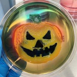
🔝Intricate chiral patterns formed by Paenibacillus dendritiformis grown on minimal media in a Petri dish